Clinical trials of interest to the Lugano dental practice
Ceramic veneers
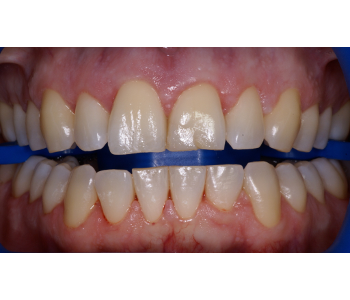
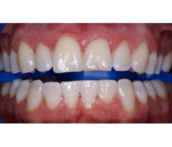
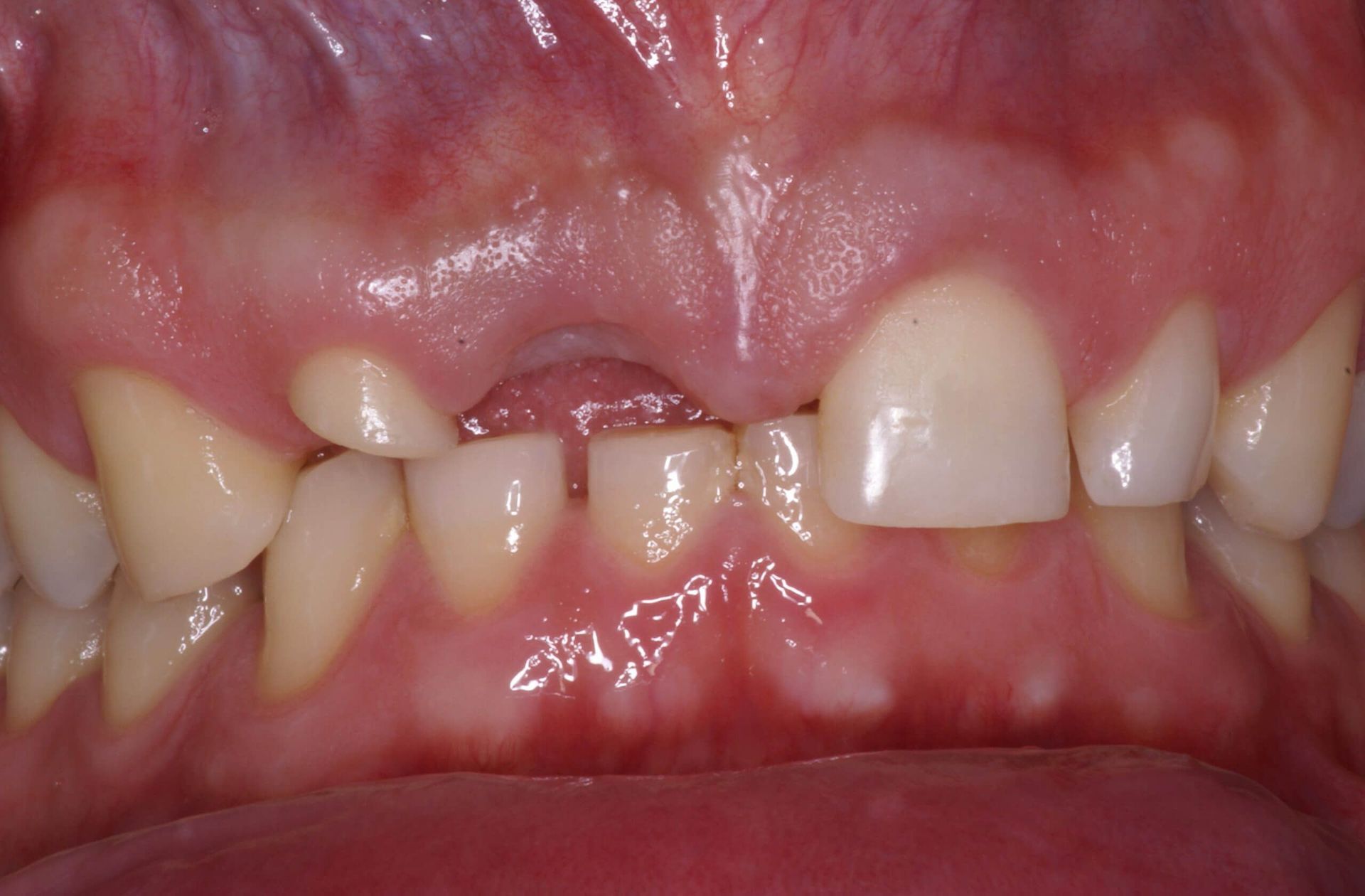
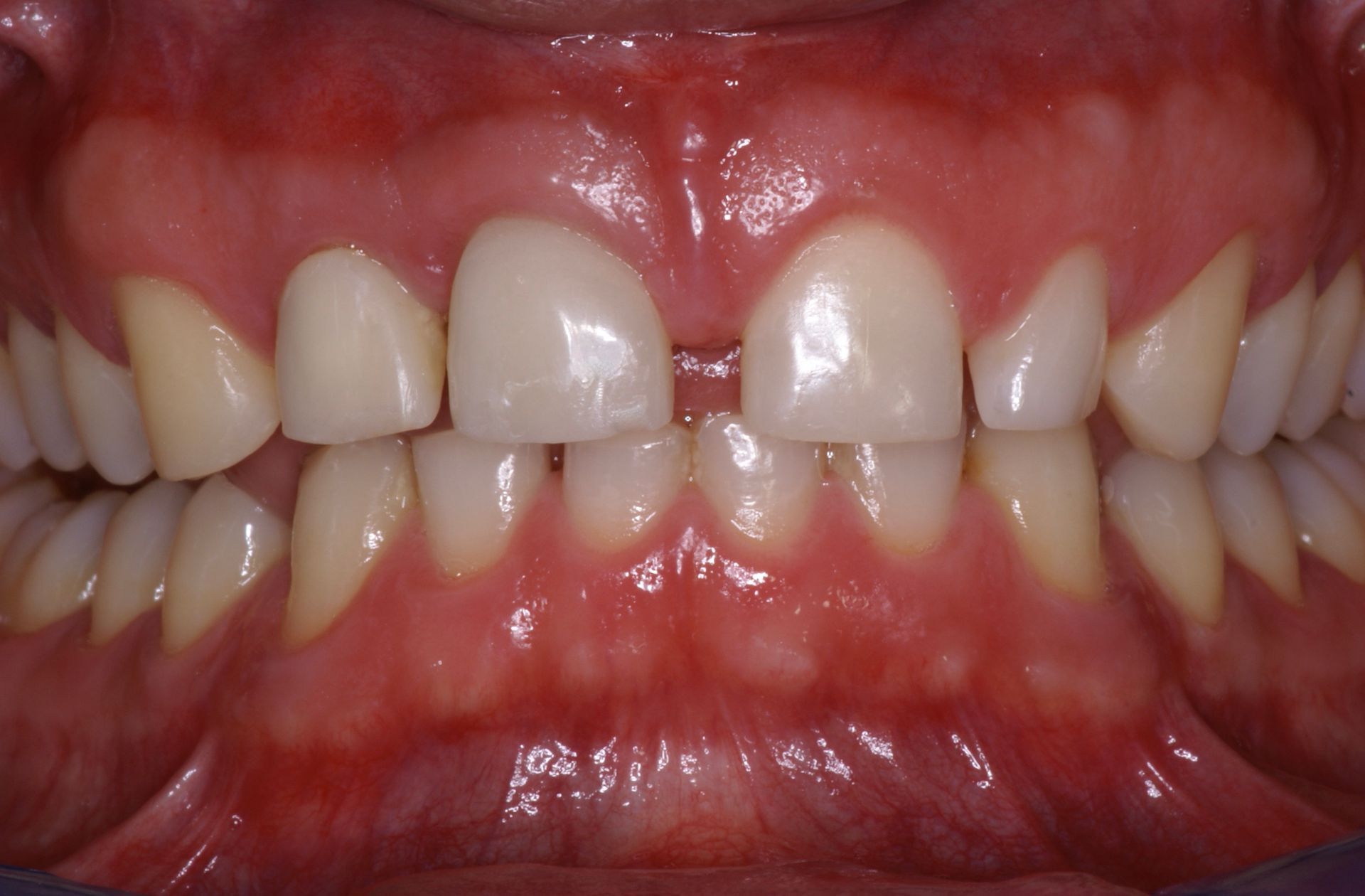
First case
The patient had an ill-fitting ceramic and metal crown on tooth 21, which caused constant inflammation of the gingiva.
In addition, teeth 11, 12 and 13 had suffered significant wear, compromising the aesthetics of the smile. The defects were corrected by applying ceramic veneers on teeth 13, 12, 11, 22 and 23, and a ceramic crown on tooth 21. This also reduced the diastema between the two central incisors.
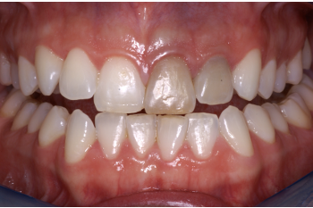
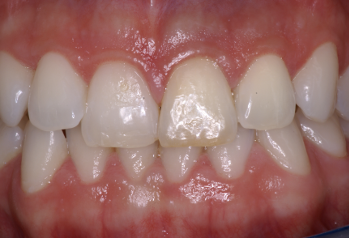
Second case
Teeth 12, 11, 21 and 22 had composite veneers that over the years had slowly degraded and no longer met the aesthetic needs of the patient.
After making a mock-up (pre-visualisation of the final result directly in the mouth) and discussing the details with the patient, we made six ceramic veneers. The products allowed the interdental spaces to be closed slightly, creating a brilliant smile and rejuvenating the appearance of the face.
Third case
The patient wanted to improve the appearance of the smile as the upper incisors 11 and 21 were divergent and 12 and 22 were cone teeth (smaller than normal).
The orthodontic treatment proposed by us allowed the two central incisors to be repositioned to a more natural position; the lateral incisors were covered with ceramic veneers to improve shape and size.
Zoom 2 whitening
Teeth whitening was achieved by applying a hydrogen peroxide gel to the teeth and amplifying its effect through light activation with the ZOOM 2 lamp. The results are always very satisfying, creating a Hollywood smile.
Internal teeth whitening
The patient suffered a dental trauma at a young age that affected teeth 21 and 22, which later became extremely dark. The solution was to proceed with internal tooth whitening. The patient was very satisfied with the result and did not want other treatments.
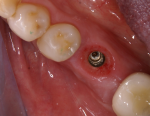
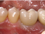
Simple case of implantology
The patient suffered the loss of the first left lower molar (36) due to a root fracture. Since the adjacent teeth were in excellent condition, it was decided to replace the tooth with a Straumann bone-level implant. After a sufficient period of time, the impression was taken and a ceramic tooth was made and screwed to the implant. The access channel to the occlusal screw was then closed with an aesthetic filling. It is possible to see the perfect integration of the item and the presence of the gingival papillae that close the interdental spaces.
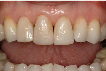
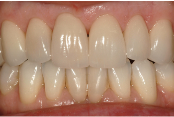
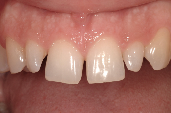
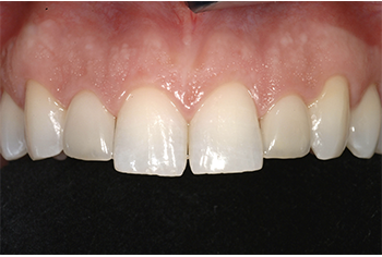
Complex cases of implantology and fixed prosthesis
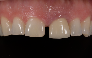
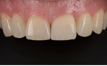
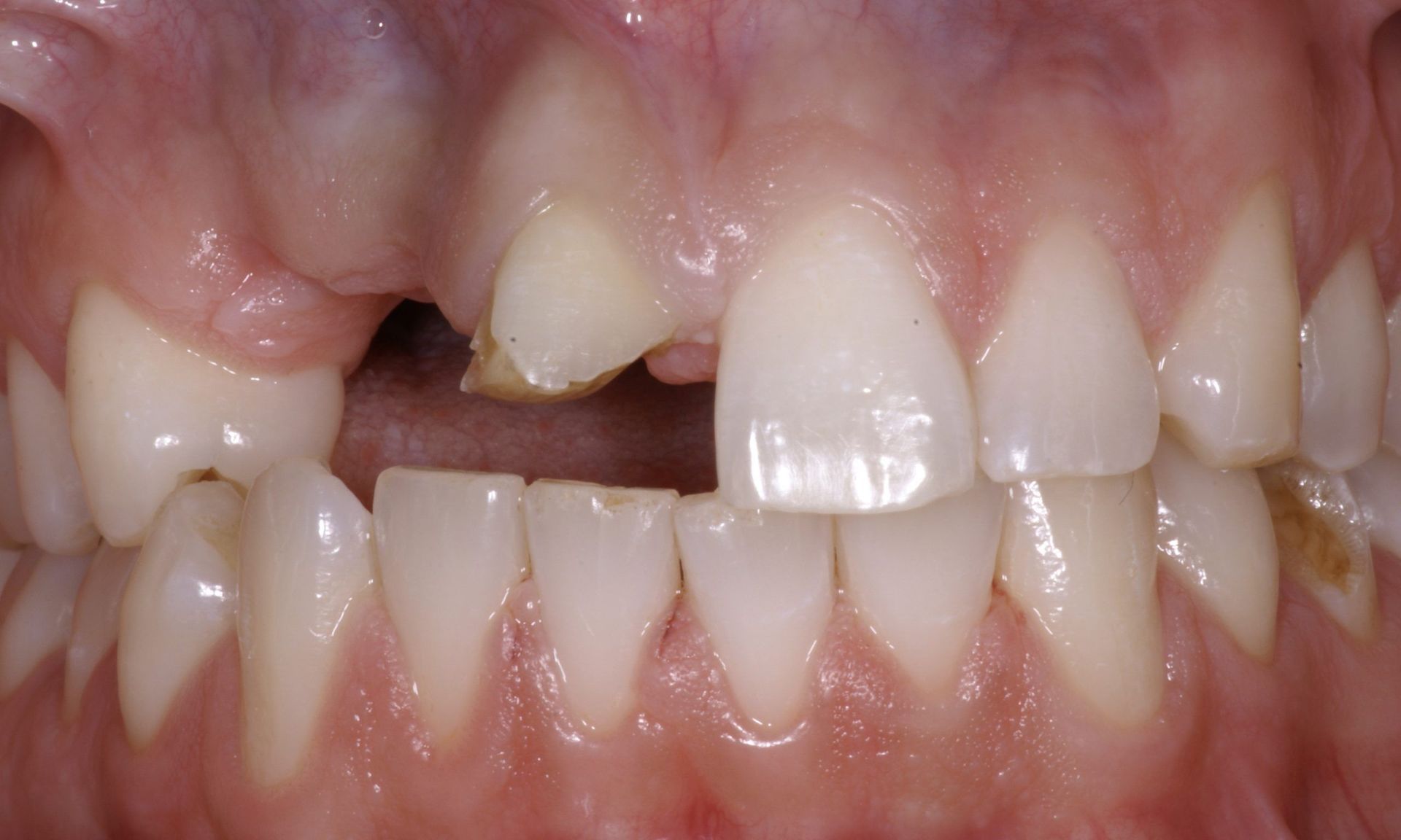
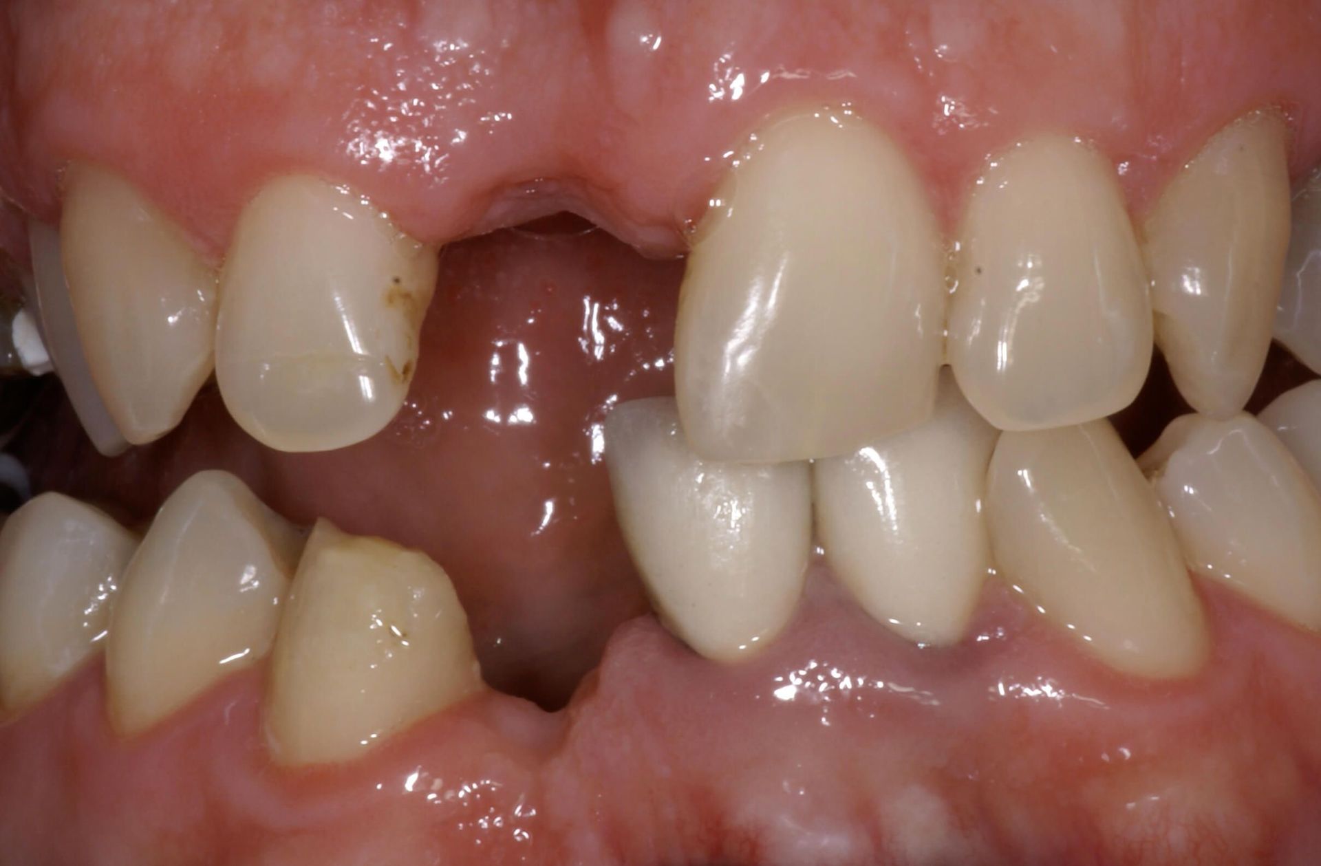
First case
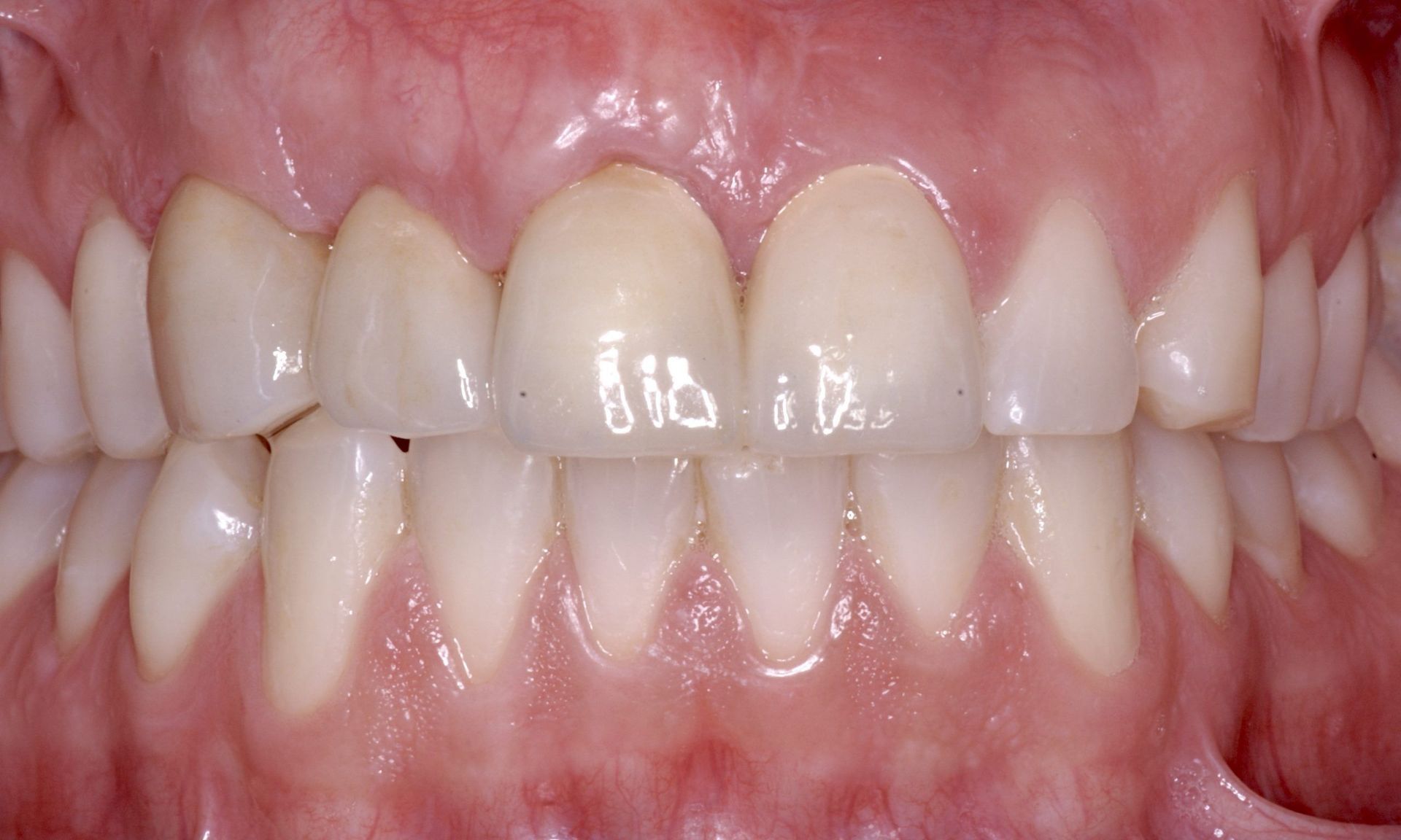
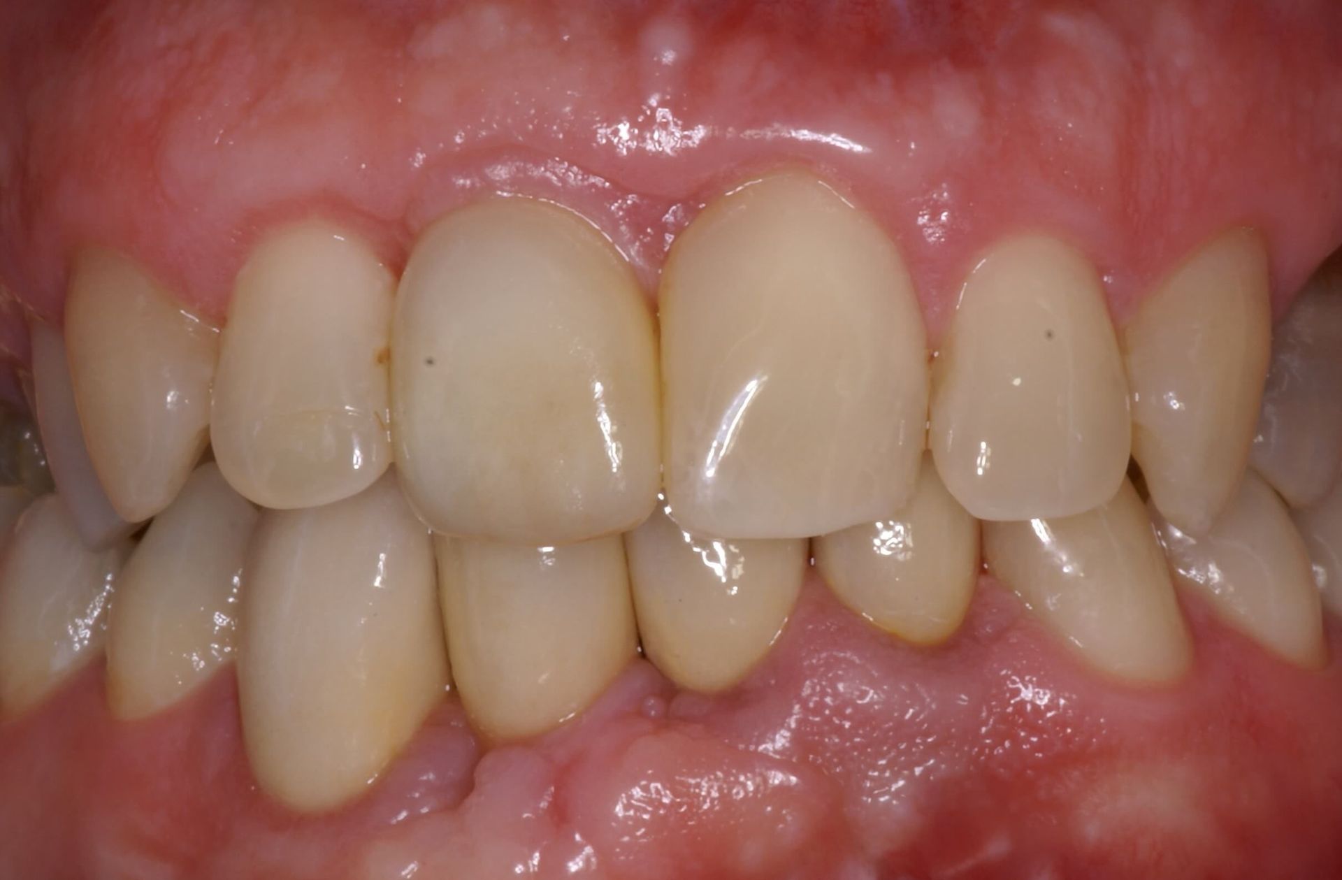
The patient suffered a severe trauma at work that resulted in a sharp fracture of the central incisor (11) and a partial fracture of the lateral incisor (12). The latter was saved by performing a root canal and then covering with a ceramic crown. Tooth 11 had to be extracted and, after a period of healing, a Straumann implant was placed and reconstructed with a ceramic crown.
Second case
The patient suffered two injuries a few months apart that resulted in the fracture of the central incisors, the loss of the canine, lateral incisor and vestibular bone table. In order to insert an implant in the canine area in order to replace the two missing teeth, a bone transplant was necessary. After several months of waiting, which allowed a good integration of the graft, a bone-level Straumann implant was inserted. The final prosthetic phase was realised with a small extension bridge over the canine and ceramic crowns.
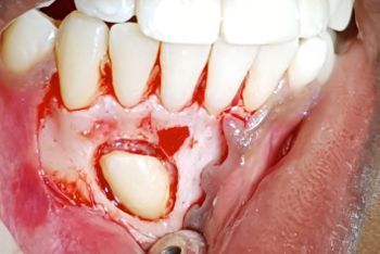
Third case
As a result of an accident, the patient lost a lower incisor, fractured the canine and lost an upper incisor along with its vestibular bone plate.
It was necessary to perform a bone graft on the upper arch before placing a bone-level Straumann implant. The same type of implant was placed on the lower arch and a small bone augmentation was performed at the same time.
In order to harmonise the colour of the restoration and improve the aesthetics, the old crowns of the lower incisors were replaced in the final prosthetic rehabilitation.
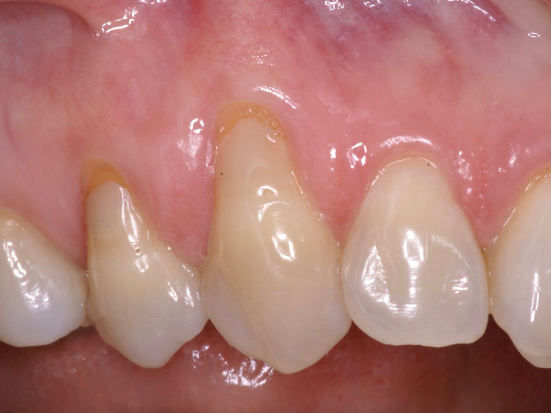
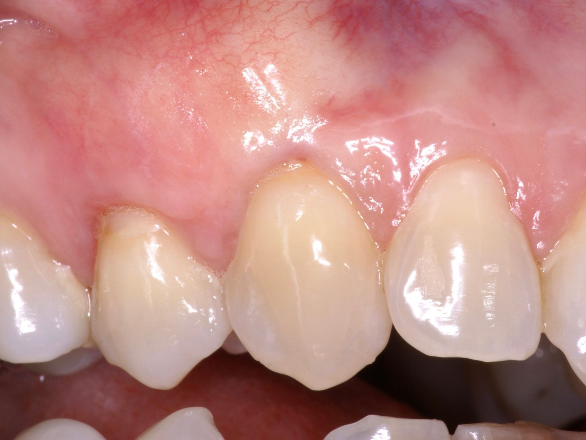
Periodontal surgery: neck coverage
First case
The presence of gingival recession at the canine neck and upper premolar was very significant and unsightly. The pericoronal tissue was therefore very thin with little attached gingiva.
In order to prevent excessive recession that could jeopardise the periodontal stability of the canine, a coronal repositioning flap was created with the insertion of connective tissue taken from the palate. The final result was very satisfactory and ensured greater resistance of the tissue to mechanical irritation.
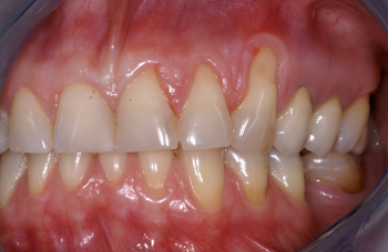
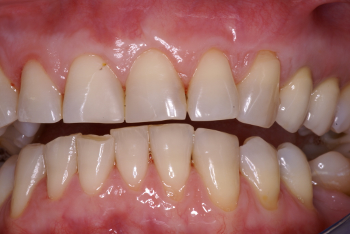
Second case
A patient had very significant gum recession on teeth 21, 22 and 23 caused by incorrect use of the toothbrush.
In this case the patient was first instructed about suitable brushing technique and, later, a coronal gingival repositioning was carried out with the addition of a graft of connective tissue taken from the palate area. The thickening of the gum adhering around the tooth neck allowed a stable result to be maintained in the long term.
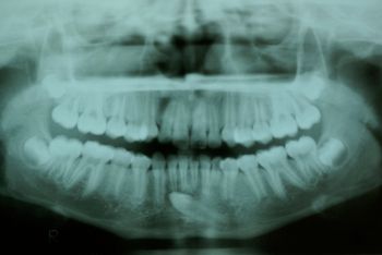
Included tooth surgery
In this case, the patient had an included lower left canine in a position that did not allow its alignment on the dental arch by orthodontic treatment. Surgical extraction of the tooth was therefore performed.